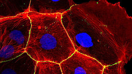
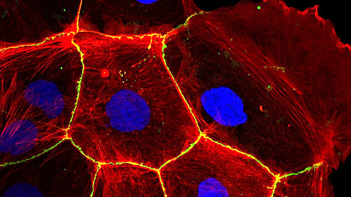

The research group headed by Prof. Dr. A. Ngezahayo conducts research in the field of cell-cell communication in connection with diseases such as cancer or stroke as well as with organ development and function such as brain, heart or bone.
The research group headed by Prof. Dr. A. Ngezahayo conducts research in the field of cell-cell communication in connection with diseases such as cancer or stroke as well as with organ development and function such as brain, heart or bone.
Oligomerisation of connexin in connexin channels
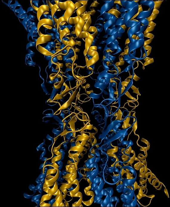
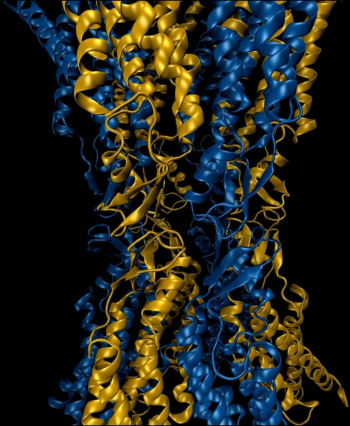
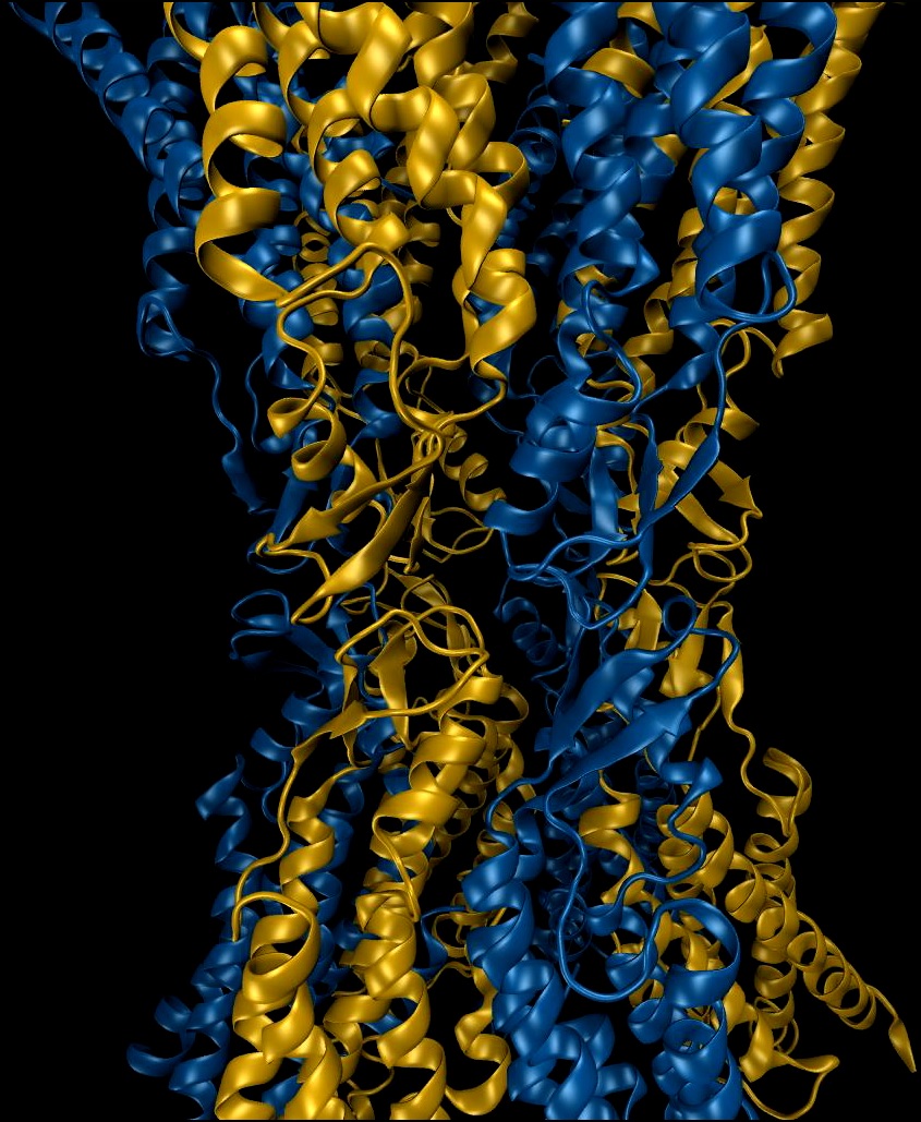
Connexins are membrane proteins that hexamerize and form channels in the cell membrane. These channels, when opened, are large enough to allow the exchange of metabolites between the intra- and extracellular spaces. The channels in adjacent cells can dock to each other and form gap junction channels. They allow then an exchange of information in form of ions and metabolites such as second messengers. The connexin channels as hemichannels or gap junction channels, lead to synchronization of cell function in tissue e.g. the synchronized contraction of cardiomyocytes. Modification of the connexin interaction and hemichannel docking surfaces, as results of genetic mutations, are related to various diseases. The analysis of the mutants in comparison to the wild type allows to understand the formation of stable functional cell to cell tunnels.
Metabolite transporters in the cell membrane
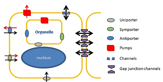
Connexin channels represent metabolite transporters between cells. The cells have also other metabolite transporters such as ion channels. Those transporters are found in all living systems and serve as uptake for different elements by facilitating their diffusion trough the cell membrane. In eukaryotic cells, the transporters are involved in tissue development and function such as generator of electrical or Ca2+ signals. Our group applies electrophysiological as well as imaging methods to analyze how those transporters are regulated at the molecular level.
Integration of connexin channels in general cell signaling
Involvement in tissue pathophysiology
For a proper function of tissue, connexin channels must be integrated in general signaling in tissue. In our group we analyze the integration of connexin channels in the purinergic signaling. The purinergic signaling is composed of two types of receptors: P1 receptors which bind adenosine, and P2 receptors which bind the nucleotides ATP, ADP and UTP. Proteomic data proposed to consider the purinergic signaling and the Cx channels as an integrated functional unit. This integrated functional unit is essential for the development and function of tissue such as vascular system, brain or epithelia. Our group participate in the elucidation of the molecular mechanisms that allow the integration of connexin channels in the purinergic tissue in various tissues.
Interaction between connexin channels and epithelial barrier function
Epithelia e.g. pulmonary epithelium maintain a barrier that separates the atmospheric space (luminal) from the body circulatory system lying beneath (abluminal). For the formation of the barrier, epithelial cells express claudins and occludins that form tight junctions, the morphological gasket-like seals that encircle epithelial cells around the apical pole. The tight junctions close the intercellular spaces and regulate thereby the paracellular diffusion. They also separate the apical pole from the basolateral cellular region, allowing the epithelial cells to maintain their polarity. Pathophysiological reactions in tissue, such as inflammation, induce locally liberated mediators that perturb the barrier function of the epithelium. Suggesting that the mediator induced information spreads in the tissue, connexin channels play a role. We develop methods to analyze the interaction between connexin channels and the tight junctions.
Cell cultures in 3D systems to simulate tissue conditions
Working in cell cultures and expression systems give valuable information. However, an original tissue function as 3D system containing different cell types. We are engaged in developing a 3D culture system that should allow us to follow the physiology of the cells and their interactions in a tissue-like system. Moreover we would be able to simulate pathological events such as inflammation or even development of pathology such as neoplastic transformation to test diagnostic and even therapeutic interventions. These 3D systems will not completely replace but will strongly reduce the animal experimentatins.
Contact
30419 Hannover


